Works
Deep Auto Search
deep learning
Introduction
To validate scientific findings, a large amount of data is essential. However, manually collecting this data in microscopy is both tedious and time-consuming. Deep-auto-search harnesses the power of deep learning to automate the complex, repetitive tasks in microscopy, effectively taking over from humans. This technology autonomously captures high-quality cell images. In this work, data collection was fully automated, integrating my projects on cell segmentation , which centers cells in the field of view (FOV), and key frame detection, which identifies the optimal focal plane.
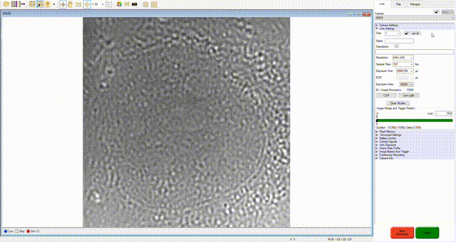
Demonstration
This demo speeds up moving cells to the center, recording, and saving by 15 times, and finds the desired focal plane 0.8 times slower. The operation is achieved by integrating the light source, shutter, stage, and camera.
Human behavior on microscope:
- Stage adjustment: identify the object and move it to the center of the field of view
- Focus scanning: adjust the focal plane back and forth to ensure features are sharp and clear
- Quality control: record if the quality of cell meets the demand
How machine can reproduce the operation
- Segmentation: define outlines around cells to separate them from the background
- Auto focus: adjust the height to obtain the highest-quality images from a set of cell images at different focal planes
- Binary classification: verify that the quality of the cell image meets human expectations
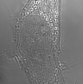
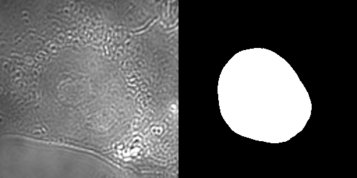
Cell Segmentation
The Eff-UNet model converts raw images into a binary mask that highlights the cell. This mask helps the machine locate the cell within the image
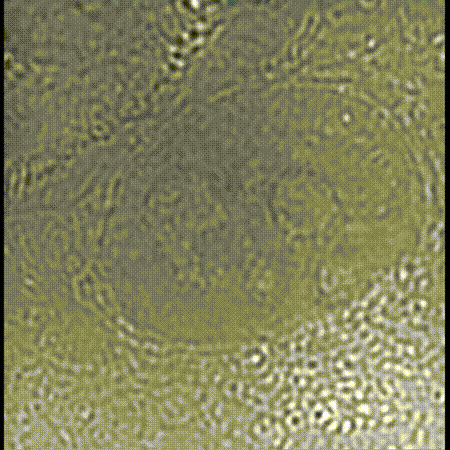
Autofocus
In microscopy, keeping objects in focus is crucial. Over time, the stage can shift, causing both sideways and up-and-down drift. Automated machine correction is essential for maintaining focus during long measurements.
Result
Cells were automatically captured in images over three hours. A separate machine, trained to classify images, ensured quality control, eliminating the need for manual review.